PDB-code

| Kinase structure | |
| Kinase: | PI4K2B |
| Family: | PIK |
| Group: | Atypical |
| Species: | HUMAN |
| IUPHAR/BPS ID: | 2499 |
| PDB: | 8A5X |
| sc-PDB: | |
| KIDFamMap: | Search |
| Release date: | 2022-10-05 |
| PubMed: | 36184029 |
| Chain: | A |
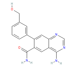
| Orthosteric ligand: | L6A |
 | |

| Structural information | |
| DFG conformation: | out-like |
| αC-helix conformation: | in |
| G-rich loop angle (distance): | 54.6° (17Å) |
| G-rich loop rotation: | 36.9° |
| Quality Score: | 7.6 |
| Resolution: | 2.4 Å |
| Missing Residues: | 0 |
| Missing Atoms: | 0 |
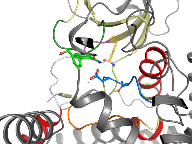
| Ligand binding mode | ||||
| Pockets | Subpockets | |||
| front | ||||
| Pocket alignment | |
| Uniprot sequence: | ERISQGSSGSYFVGVFKPKGYLSEAGAYLVDNSIVPKTKVVGSFQLFVEYKEAEYWLRKLDYI--IRNTDRGNDNWLVAIDNGLA |
| Sequence structure: | ERISQGSSGSYFVGVFKPKGYLSEAGAYLVDNSIVPKTKVVGSFQLFVEYKEAEYWLRKLDYI__IRNTDRGNDNWLVAIDNGLA |
| Ligand affinity | |
| Ligand not found in ChEMBL. | |
Kinase-ligand interaction pattern
• Hydrophobic • Aromatic face-to-face • Aromatic face-to-edge • H-bond donor • H-bond acceptor • Ionic positive • Ionic negative
| I | g.l | II | III | αC | |||||||||||||||
| 1 E 124 | 2 R 125 | 3 I 126 | 4 S 127 | 5 Q 128 | 6 G 129 | 7 S 130 | 8 S 131 | 9 G 132 | 10 S 133 | 11 Y 134 | 12 F 135 | 13 V 136 | 14 G 145 | 15 V 146 | 16 F 147 | 17 K 148 | 18 P 149 | 19 K 150 | 20 G 185 |
| • | • | • | •• | ||||||||||||||||
| αC | b.l | IV | |||||||||||||||||
| 21 Y 186 | 22 L 187 | 23 S 188 | 24 E 189 | 25 A 190 | 26 G 191 | 27 A 192 | 28 Y 193 | 29 L 194 | 30 V 195 | 31 D 196 | 32 N 197 | 33 S 202 | 34 I 203 | 35 V 204 | 36 P 205 | 37 K 206 | 38 T 207 | 39 K 208 | 40 V 209 |
| IV | V | GK | hinge | linker | αD | αE | |||||||||||||
| 41 V 210 | 42 G 254 | 43 S 255 | 44 F 256 | 45 Q 257 | 46 L 258 | 47 F 259 | 48 V 260 | 49 E 261 | 50 Y 263 | 51 K 264 | 52 E 265 | 53 A 266 | 54 E 267 | 55 Y 268 | 56 W 269 | 57 L 270 | 58 R 271 | 59 K 272 | 60 L 296 |
| •• | • | •• | •• | ||||||||||||||||
| αE | VI | c.l | VII | VIII | x | ||||||||||||||
| 61 D 297 | 62 Y 298 | 63 I 299 | 64 _ _ | 65 _ _ | 66 I 300 | 67 R 301 | 68 N 302 | 69 T 303 | 70 D 304 | 71 R 305 | 72 G 306 | 73 N 307 | 74 D 308 | 75 N 309 | 76 W 310 | 77 L 311 | 78 V 312 | 79 A 342 | 80 I 343 |
| • | |||||||||||||||||||
| DFG | a.l | ||||||||||||||||||
| 81 D 344 | 82 N 345 | 83 G 346 | 84 L 347 | 85 A 348 | |||||||||||||||
| • | |||||||||||||||||||
Interaction pattern search
Search KLIFS for kinase-ligand complexes with similar interaction patterns: