PDB-code

| Kinase structure | |
| Kinase: | PTK6 (BRK) |
| Family: | Src |
| Group: | TK |
| Species: | HUMAN |
| IUPHAR/BPS ID: | 2182 |
| PDB: | 5D7V |
| sc-PDB: | |
| KIDFamMap: | Search |
| Release date: | 2016-08-17 |
| PubMed: | 27480927 |
| Chain: | D |
| (Alternate) Model: | B |

| More entries for 5D7V | |||||
| 5D7V | Alternative model: | A | Chain: | A | |
| 5D7V | Alternative model: | A | Chain: | B | |
| 5D7V | Alternative model: | A | Chain: | C | |
| 5D7V | Alternative model: | A | Chain: | D | |
| 5D7V | Alternative model: | B | Chain: | A | |
| 5D7V | Alternative model: | B | Chain: | B | |
| 5D7V | Alternative model: | B | Chain: | C | |
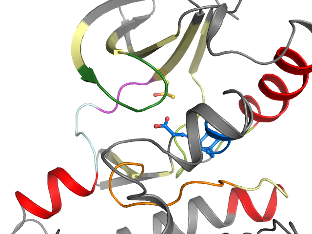
| Structural information | |
| DFG conformation: | in |
| αC-helix conformation: | out |
| G-rich loop angle (distance): | 49.5° (15.3Å) |
| G-rich loop rotation: | 66.5° |
| Quality Score: | 8 |
| Resolution: | 2.33 Å |
| Missing Residues: | 0 |
| Missing Atoms: | 0 |
| Ligand binding mode | ||||
| Waters | ||||
| Cluster I4 | Ligand No | Protein No | ||
| Pocket alignment | |
| Uniprot sequence: | RKLGSGYFGEVFEVAIKVIMLQSEIQAMKKLRKHILALYAVYIITELMAKGSLLELLRDYLESQNYIHRDLAARNILVVGDFGLA |
| Sequence structure: | RKLGSGYFGEVFEVAIKVIMLQSEIQAMKKLRKHILALYAVYIITELMAKGSLLELLRDYLESQNYIHRDLAARNILVVGDFGLA |