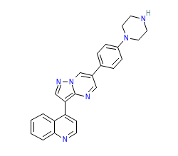
PDB-code
| More entries for 3MDY | |||||
| 3MDY | Alternative model: | A | Chain: | A | |
| 3MDY | Alternative model: | A | Chain: | C | |
| 3MDY | Alternative model: | B | Chain: | A | |
| Structural information | |
| DFG conformation: | in |
| αC-helix conformation: | in |
| G-rich loop angle (distance): | 47.2° (13.8Å) |
| G-rich loop rotation: | 79.1° |
| Quality Score: | 9.6 |
| Resolution: | 2.05 Å |
| Missing Residues: | 0 |
| Missing Atoms: | 4 |
| Ligand binding mode | ||||
| Pockets | Subpockets | Waters | ||
| front | FP-I | Cluster I2 I5 | Ligand No Yes | Protein Yes Yes |
| Pocket alignment | |
| Uniprot sequence: | KQIGKGRYGEVWMVAVKVFSWFRETEIYQTVLENILGFIAAYLITDYHENGSLYDYLKSTQGKPAIAHRDLKSKNILVIADLGLA |
| Sequence structure: | KQIGKGRYGEVWMVAVKVFSWFRETEIYQTVLENILGFIAAYLITDYHENGSLYDYLKSTQGKPAIAHRDLKSKNILVIADLGLA |
| Ligand affinity | |
| ChEMBL ID: | CHEMBL513147 |
| Bioaffinities: | 19 records for 11 kinases |
| Species | Kinase (ChEMBL naming) | Median | Min | Max | Type | Records |
|---|---|---|---|---|---|---|
| Homo sapiens | Activin receptor type-1 | 7.8 | 7.4 | 8.5 | pIC50 | 3 |
| Mus musculus | Activin receptor type-1 | 7.9 | 7.9 | 7.9 | pEC50 | 1 |
| Homo sapiens | Activin receptor type-1B | 5.7 | 5.7 | 7.4 | pIC50 | 2 |
| Homo sapiens | AMP-activated protein kinase, beta-1 subunit | 6 | 6 | 6 | pIC50 | 1 |
| Homo sapiens | Bone morphogenetic protein receptor type-1A | 7.7 | 7.7 | 8.7 | pIC50 | 2 |
| Homo sapiens | Bone morphogenetic protein receptor type-1B | 7.2 | 7.2 | 9 | pIC50 | 2 |
| Homo sapiens | Bone morphogenetic protein receptor type-2 | 5.4 | 5.4 | 5.4 | pIC50 | 1 |
| Homo sapiens | Serine/threonine-protein kinase receptor R3 | 7.9 | 7.9 | 8 | pIC50 | 2 |
| Homo sapiens | TGF-beta receptor type I | 6.3 | 6.3 | 7.6 | pIC50 | 3 |
| Homo sapiens | TGF-beta receptor type II | 6.9 | 6.9 | 6.9 | pIC50 | 1 |
| Homo sapiens | Vascular endothelial growth factor receptor 2 | 6.7 | 6.7 | 6.7 | pIC50 | 1 |
Kinase-ligand interaction pattern
• Hydrophobic • Aromatic face-to-face • Aromatic face-to-edge • H-bond donor • H-bond acceptor • Ionic positive • Ionic negative
| I | g.l | II | III | αC | |||||||||||||||
| 1 K 208 | 2 Q 209 | 3 I 210 | 4 G 211 | 5 K 212 | 6 G 213 | 7 R 214 | 8 Y 215 | 9 G 216 | 10 E 217 | 11 V 218 | 12 W 219 | 13 M 220 | 14 V 228 | 15 A 229 | 16 V 230 | 17 K 231 | 18 V 232 | 19 F 233 | 20 S 240 |
| • | • | • | • | ||||||||||||||||
| αC | b.l | IV | |||||||||||||||||
| 21 W 241 | 22 F 242 | 23 R 243 | 24 E 244 | 25 T 245 | 26 E 246 | 27 I 247 | 28 Y 248 | 29 Q 249 | 30 T 250 | 31 V 251 | 32 L 252 | 33 E 256 | 34 N 257 | 35 I 258 | 36 L 259 | 37 G 260 | 38 F 261 | 39 I 262 | 40 A 263 |
| • | |||||||||||||||||||
| IV | V | GK | hinge | linker | αD | αE | |||||||||||||
| 41 A 264 | 42 Y 276 | 43 L 277 | 44 I 278 | 45 T 279 | 46 D 280 | 47 Y 281 | 48 H 282 | 49 E 283 | 50 N 284 | 51 G 285 | 52 S 286 | 53 L 287 | 54 Y 288 | 55 D 289 | 56 Y 290 | 57 L 291 | 58 K 292 | 59 S 293 | 60 T 322 |
| • | • | •• | •• | • | • | • | • | ||||||||||||
| αE | VI | c.l | VII | VIII | x | ||||||||||||||
| 61 Q 323 | 62 G 324 | 63 K 325 | 64 P 326 | 65 A 327 | 66 I 328 | 67 A 329 | 68 H 330 | 69 R 331 | 70 D 332 | 71 L 333 | 72 K 334 | 73 S 335 | 74 K 336 | 75 N 337 | 76 I 338 | 77 L 339 | 78 V 340 | 79 I 348 | 80 A 349 |
| • | • | • | |||||||||||||||||
| DFG | a.l | ||||||||||||||||||
| 81 D 350 | 82 L 351 | 83 G 352 | 84 L 353 | 85 A 354 | |||||||||||||||
| • | |||||||||||||||||||
Interaction pattern search
Search KLIFS for kinase-ligand complexes with similar interaction patterns:


